


Jörg Barner
JPK Instruments AG, Germany
Title: Mechanical and topographical characterization of biomaterials, cells and tissues by enhanced atomic force microscopy
Biography
Biography: Jörg Barner
Abstract
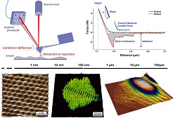
Besides structural and physico-chemical composition, the topography, roughness, adhesiveness and mechanical properties of biomaterials are relevant parameters which strongly affect cell differentiation and tissue formation, and are thus crucial for assessing biocompatibility in the human body. We have developed a multipurpose AFM device which allows comprehensive characterization of these properties and interactions on the nanoscale under physiological conditions and in combination with advanced optical microscopy. Our unique quantitative imaging (QI™) mode determines several sample properties, like topography and the Young’s modulus, with one measurement. With the CellHesion® technique, the adhesion of a single cell to any substrate can be measured and validated. The NanoWizard® ULTRA Speed technique enables fast AFM imaging of dynamic processes with approx. 1 frame per second. Using QI™, we have characterized the topography and mechanical properties of challenging samples like living cells and tissue sections. The adhesion of single fibroblast cells to various surface modifications could be quantified and their suitability as cochlear implant coatings could be assessed. Fast AFM imaging revealed collagen type I fibrillogenesis and the formation of the 67 nm D-banding in situ with high spatio-temporal resolution. Here, we are presenting an enhanced AFM, making this technique a valuable tool for biomedical research.
Recent Publications
- N. Ardjomandi, J. Huth, D. STAMOV, A. Henrich, C. Klein, H.-P. Wendel, S. Reinert, D. Alexander, 2016, “Surface biofunctionalisation of ß-TCP blocks using aptamer 74 for bone tissue engineering”, Mater Sci Eng C Mater Biol Appl 67, 267-275.
- D. R. STAMOV, S. B. Kaemmer, A. Hermsdörfer, J. Barner, T. Jähnke, H. Haschke, 2015, “BioScience AFM - Capturing dynamics from single molecules to living cells", Microscopy Today 23 (6), 18-25.
- C. Gonnermann, C. Huang, S. F. S. Becker, D. R. STAMOV, D. Wedlich, J. Kashef, C. M. Franz, 2015, “Quantitating membrane bleb stiffness using AFM force spectroscopy and an optical sideview setup”, Integr Biol 7 (3), 356-363.
- D. R. STAMOV, E. Stock, C. M. Franz, T. Jähnke, H. Haschke, 2015, “Imaging collagen type I fibrillogenesis with high spatiotemporal resolution”, Ultramicroscopy 149, 86-94.
- M. Greiner, M. Jäckel, A. C. Scheiwe, D. R. STAMOW, T. J. Autenrieth, J. Lahann, C. M. Franz, M. Bastmeyer, 2014, “Multifunctional polymer scaffolds with adjustable pore size and chemoattractant gradients for studying cell matrix invasion”, Biomaterials 35 (2), 611-619.